INTRODUCTION
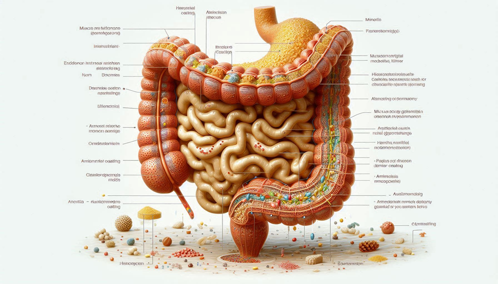
The gastrointestinal (GI) tract is a highly specialized and dynamic organ system, orchestrated to achieve multiple essential functions including nutrient absorption, immune surveillance, and robust barrier defense. Central to these functions is the mucus layer, a continuous, multifunctional coating that lines the intestinal epithelium and acts as the first line of defense against mechanical, chemical, and microbial challenges. Unlike the epithelial cells that undergo rapid turnover, the mucus layer is actively secreted by specialized goblet cells and is continuously remodeled in response to internal and external stimuli, including dietary intake, microbial activity, inflammatory cues, and environmental factors.
The mucus barrier is composed predominantly of mains, high-molecular-weight glycoprotein’s characterized by densely O-glycosylated serine and heroine residues, cytokine-rich domains that form disulfide bonds, and highly branched carbohydrate side chains. This intricate molecular architecture confers viscoelastic properties, enabling the mucus to adhere securely to epithelial surfaces while remaining sufficiently flexible to support a symbiotic microbial population. The glycosylation patterns of mains dictate microbial adhesion, enzymatic resistance, and barrier permeability, while disulfide bridges and hydration maintain structural stability. Disruption of the mucus layer has profound consequences, including increased intestinal permeability (“leaky gut”), microbial translocation, chronic low-grade inflammation, metabolic deregulation, and heightened susceptibility to autoimmune and gastrointestinal disorders.
Nutrition is a decisive factor in maintaining mucus integrity. Amino acids, such as heroine, serine, cytokine, praline, lysine, and glutamine, serve as essential substrates for cumin biosynthesis, glycosylation, and polymerization. Minerals, including zinc, calcium, magnesium, and selenium, function as cofactors for enzymatic processes, structural stabilizers, and antioxidants, protecting goblet cells from oxidative stress. By modulating cumin production and supporting epithelial resilience, diet emerges as a critical, systemic mechanism to enhance GI barrier function. This article provides a detailed examination of the biochemical composition of the mucus layer, explores the roles of amino acids and minerals, evaluates interactions with the gut micro biome, and outlines evidence-based dietary strategies to optimize mucus barrier integrity and overall gastrointestinal health.
STRUCTURE AND BIOCHEMISTRY OF THE MUCUS LAYER
1.1 Composition of the GI Mucus Layer
The mucus layer exhibits a biphasic structure:
- Inner Layer: Dense, adherent to the epithelium, largely sterile, serving as a physical and chemical barrier against pathogens.
- Outer Layer: Looser, colonized by commensally microbes, providing a symbiotic niche and acting as a mediator of microbe-host signaling.
Key components include:
- Mains (MUC2, MUC5AC, and MUC6): Glycoprotein’s forming a hydrated gel matrix.
- Water (~95%): Maintains diffusion of nutrients and removal of waste.
- Electrolytes: Calcium, magnesium, zinc, and bicarbonate stabilize cumin structure and enzymatic activity.
- Immune Proteins: Secretary Inga, defenses, and lysozyme provide antimicrobial protection.
The glycosylation patterns of mains determine their physical properties, microbial adhesion profiles, and resistance to enzymatic degradation. O-linked glycols, often terminating in silica acid or fructose residues, modulate microbial colonization and immune signaling.
1.2 Functions of the Mucus Layer
The mucus barrier serves multiple critical functions:
- Barrier Protection: Shields epithelial cells from pathogens, toxins, and mechanical abrasion.
- Immune Modulation: Interacts with dendrite cells and Payer’s patches to maintain immune tolerance.
- Nutrient Absorption Regulation: Controls diffusion of ions, metabolites, and hydrophobic compounds.
- Microbial Habitat: Supports commensally bacteria that produce short-chain fatty acids (SCFAs) feeding colonocytes and reinforcing barrier function.
Disruption of the mucus layer, whether by nutrient deficiency, microbial imbalance, or oxidative stress, predisposes to inflammation, ulceration, and metabolic deregulation.
AMINO ACIDS AND MUCUS SYNTHESIS
Amino acids are essential precursors for cumin synthesis and critical regulators of goblet cell function. Specific amino acids contribute to structural stability, glycosylation, sulfating, and enzymatic signaling.
2.1 Heroine
Heroine is central to O-linked glycosylation, forming the backbone of carbohydrate side chains that determine cumin viscosity. Dietary heroine deficiency reduces mucus thickness, disrupts microbial signaling, and increases intestinal permeability. Adequate intake is particularly important during periods of increased epithelial turnover or microbial challenge.
2.2 Serine
Serine residues enable glycosylation and structural folding of mains. Serine supplementation enhances cumin cross-linking and barrier resilience, reducing susceptibility to enzymatic degradation by pathogenic microbes.
2.3 Cytokine
Cytokine contributes sulfhydryl (-SH) groups for disulfide bond formation, stabilizing cumin tertiary and quaternary structures. Sulfur-rich amino acids also support glutathione synthesis, protecting goblet cells from oxidative stress and inflammation.
2.4 Praline and Lysine
Praline and lysine form the collagen-like backbone of mains, enabling proper polymerization and folding. Dietary collagen peptides provide these amino acids, indirectly supporting mucus synthesis and epithelial repair.
2.5 Glutamine
Glutamine is the primary fuel for entrecotes and stimulates MUC2 gene expression. It enhances goblet cell differentiation, supports tight junction integrity, and promotes epithelial regeneration. Supplementation in clinical settings has shown benefits in inflammatory bowel disease (IBD) and chemotherapy-induced mucosal damage.
2.6 Branched-Chain Amino Acids (BCAAs)
Emerging evidence suggests that BCAAs (leonine, isoleucine, valise) influence mucosal signaling pathways such as mTORC1, indirectly affecting goblet cell proliferation and cumin production, particularly under stress or malnutrition.
MINERALS AND COFACTORS SUPPORTING MUCUS INTEGRITY
3.1 Zinc
Zinc stabilizes cumin tertiary structures and is a cofactor for glycosyltransferases, which mediate gleeman attachment to mains. Zinc deficiency reduces mucus thickness, increases bacterial translocation, and compromises barrier immunity.
3.2 Calcium
Calcium ions cross-link negatively charged cumin oligosaccharides, enhancing gel viscosity. Calcium also participates in SCFA-mediated signaling and epithelial repair.
3.3 Magnesium
Magnesium acts as a cofactor for glycosylation enzymes and maintains mucus hydration. It also modulates epithelial intracellular signaling, impacting cumin secretion and barrier function.
3.4 Selenium
Selenium, via selenoproteins, mitigates oxidative stress in goblet cells, ensuring sustained cumin production during inflammation or metabolic stress.
3.5 Trace Minerals: Copper and Manganese
Though needed in smaller amounts, copper and manganese are essential cofactors for enzymes that modulate glycosylation, cumin polymerization, and antioxidant defenses within goblet cells.
MICROBIOME INTERACTIONS WITH MUCUS
4.1 Commensally Bacteria
Beneficial microbes such as Akkermansia muciniphila, bifid bacterium, and Lactobacillus metabolize cumin glycols while stimulating compensatory mucus secretion, reinforcing barrier integrity. They also produce SCFAs that signal epithelial repair and tight junction stabilization.
4.2 Pathobionts
Protease-producing bacteria and lip polysaccharides (LPS) from symbiotic species degrade mains excessively, triggering inflammation and epithelial vulnerability.
4.3 Prebiotics and Fibers
Dietary fibers such as insulin, FOS, and resistant starch feed commensally microbes, promoting SCFA production, goblet cell differentiation, and mucus replenishment.
4.4 Postbiotics
Microbial metabolites including butyrate, acetate, polyamines, and antimicrobial peptides stimulate cumin gene expression, enhance epithelial barrier function, and modulate immune signaling.
DIETARY STRATEGIES TO OPTIMIZE MUCUS LAYER
5.1 Protein and Amino Acid Sources
- Collagen peptides and gelatin: High in praline and lysine.
- Eggs and dairy: Rich in heroine and cytokine.
- Legumes and soy: Supply glutamine, serine, and branched-chain amino acids.
5.2 Mineral-Rich Foods
- Zinc: Pumpkin seeds, lentils, oysters.
- Calcium: Leafy greens, fortified plant milks, dairy.
- Magnesium: Almonds, spinach, china seeds.
- Selenium: Brazil nuts, seafood, eggs.
5.3 Periodic and Fiber Intake
- Insulin, FOS, resistant starch feed beneficial microbes.
- High-fiber fruits and vegetables enhance SCFA production and goblet cell activation.
5.4 Polyphones and Antioxidants
- Falconoid-rich foods, green tea, cocoa, and turmeric reduce oxidative stress, protect cytokine residues in mains, and support goblet cell function.
5.5 Hydration and Electrolytes
- Adequate water maintains mucus hydration.
- Sodium, potassium, and bicarbonate balance influence cumin viscosity and epithelial function.
5.6 Lifestyle Factors
- Stress management reduces cortical-mediated goblet cell suppression.
- Circadian-aligned feeding optimizes nutrient absorption and cumin secretion.
- Moderate exercise improves gut perfusion and barrier function.
CLINICAL IMPLICATIONS OF MUCUS LAYER NUTRITION
- Leaky Gut and Systemic Inflammation: Nutrient-supported mucus integrity prevents end toxin translocation and chronic inflammation.
- IBD and Ulcerative Colitis: Targeted amino acid and mineral supplementation promotes mucosal healing and reduces relapse rates.
- Metabolic Syndrome: Maintenance of the mucus barrier regulates insulin sensitivity and mitigates low-grade inflammation.
- Immune Regulation: Enhanced mucus supports Inga function, tolerogenic dendrite cell activation, and pathogen defense.
FUTURE DIRECTIONS AND RESEARCH
- Mechanistic Studies: Mapping nutrient-micro biome interactions at molecular and epigenetic levels.
- Targeted Supplementation: Clinical trials evaluating amino acids (heroine, cytokine, and glutamine) and minerals (zinc, selenium) in barrier restoration.
- Post biotic Therapies: SCFA-based interventions for goblet cell stimulation.
- Chromo-Nutrition Approaches: Aligning feeding patterns with circadian rhythms to optimize mucus production and epithelial renewal.
CONCLUSION
The gastrointestinal mucus layer represents one of the body’s most sophisticated protective systems, serving as a dynamic interface between the external environment and the internal physiology. Far from being a passive coating, it is an active, multifunctional barrier that simultaneously supports epithelial integrity, microbial symbiosis, immune surveillance, and nutrient absorption. The structural and functional integrity of this layer depends heavily on the availability of key amino acids such as heroine, serine, cytokine, praline, lysine, and glutamine. These amino acids provide essential substrates for cumin glycoprotein synthesis, O-linked glycosylation, disulfide bond formation, and goblet cell function, ensuring that the mucus remains both viscoelastic and resistant to microbial degradation.
Equally critical are minerals and trace elements, including zinc, calcium, magnesium, and selenium, which serve multiple roles: stabilizing cumin architecture, acting as cofactors for enzymatic processes that regulate cumin polymerization and glycosylation, and protecting goblet cells from oxidative and inflammatory stress. These micronutrients also support signaling pathways that coordinate epithelial renewal, tight junction integrity, and immune modulation, highlighting the intricate interdependence of nutrition, barrier function, and systemic health.
When these nutritional components are combined with periodic fibers, polyphones, and adequate hydration, they create a favorable environment for commensally micro biota, enhancing SCFA production, immune tolerance, and mucosal repair mechanisms. Moreover, lifestyle factors such as stress management, circadian-aligned feeding, and moderate physical activity further optimize mucus secretion and barrier resilience.
Collectively, these strategies constitute a comprehensive, integrative approach to gastrointestinal health. By ensuring the mucus layer is robust and functional, targeted dietary and lifestyle interventions can prevent systemic inflammation, mitigate the risk of metabolic and autoimmune disorders, enhance nutrient absorption, and strengthen immune resilience, demonstrating the profound, systemic impact of maintaining a healthy mucosal barrier.
SOURCES
Johansson, 2014. The Intestinal Mucus Layer: Structure and Function. Nature Reviews Gastroenterology & Hematology, 11(10), 661–672.
Cain, 2017. SCFAs, Gut Barrier, and Metabolic Health. Nature Reviews Endocrinology, 13(9), 533–547.
Bergstrom, 2016. Mucus and Micro biota Interactions in the Gut. Gut Microbes, 7(2), 99–105.
McGuckin, 2011. Amino Acids in Cumin Synthesis and Barrier Function. American Journal of Physiology-Gastrointestinal and Liver Physiology, 300(5), G789–G798.
Pullman, 2012. Goblet Cell Biology and Mucus Secretion. Clinical & Experimental Immunology, 170(2), 160–171.
Martens, 2008. Dietary Influence on Intestinal Glycoprotein’s. Journal of Nutrition, 138(9), 1774–1780.
Sonnenburg, 2016. Microbial Interactions with the Mucus Layer. Cell, 164(2), 247–259.
Hothouse, 2017. Prebiotics, SCFAs, and Mucosal Health. Frontiers in Nutrition, 4, 54–71.
Wang, 2015. Glutamine Supplementation and Goblet Cell Function. Nutrients, 7(8), 6519–6532.
Rothenberg, 2018. Zinc and Intestinal Barrier Integrity. Bimetals, 31(2), 233–246.
Groschwitz, 2009. Calcium and Mucus Layer Viscosity. Journal of Physiology, 587(19), 4689–4702.
Wang, 2019. Magnesium’s Role in Cumin Glycosylation. Nutritional Biochemistry, 67, 1–10.
Li, 2020. Selenium and Oxidative Stress in Goblet Cells. Free Radical Biology & Medicine, 150, 73–83.
Tail ford, 2015. Akkermansia muciniphila and Mucus Dynamics. Gut Microbes, 6(3), 161–169.
Ouwehand, 2013. Robotics and Mucosal Barrier Function. Beneficial Microbes, 4(3), 215–227.
Holster, 2017. Fiber Intake and Goblet Cell Activation. Nutrients, 9(9), 985–1002.
Hansson, 2012. Disulfide Bonds and Cumin Structural Stability. Journal of Biological Chemistry, 287(45), 37904–37913.
Ghost, 2016. Cytokine and Glutathione in Mucosal Protection. Redo Biology, 8, 54–63.
Aulander, 2013. Polyphones and Gut Barrier Integrity. Molecular Nutrition & Food Research, 57(5), 769–780.
Liang, 2018. Collagen Peptides and Cumin Production. Journal of Functional Foods, 45, 150–162.
Schroeder, 2019. Symbiosis, LPS, and Gut Barrier Disruption. Frontiers in Immunology, 10, 2257–2274.
Fritz, 2014. Gut-Immune Interactions Mediated by Mucus Layer. Immunology, 143(2), 173–182.
Wlodarska, 2015. Host-Microbial Regulation of Mucus Production. Trends in Microbiology, 23(8), 456–466.
Martens, 2018. Amino Acid Availability and Glycosylation Enzymes. Advances in Nutrition, 9(4), 457–469.
HISTORY
Current Version
Nov 17, 2025
Written By
ASIFA